FOLLOW US ON:
GET THE NEWSLETTER
CONTACT US
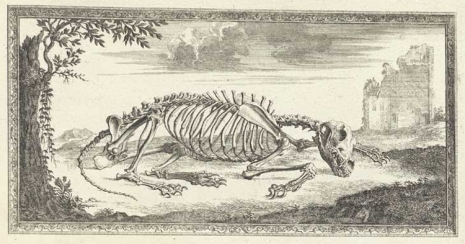
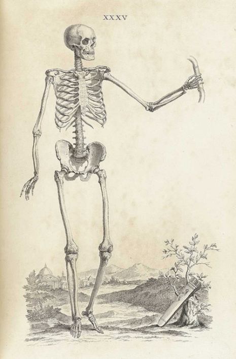
Skeletal remains: The first accurate representation of ‘The Anatomy of Bones’ from 1733
06.05.2017
10:43 am
Topics:
Tags:

014bonesskeleton14.jpg
 
Bones. This is what we come to once we’re dead and the soft tissue has gone. Bones. The sturdy architecture that shapes and protects our bodies. Most of us will end up as dust or ashes, or if very, very lucky, may one day become fossilized and exhibited in a museum as an example of a dumb 21st-century Homosapien. There’s nothing else once we’re dead. No seventy-two virgins (or is it dried fruit?), no Alleluia chorus, no wings and no harp, just the remnants of a structure that once held us together.

Humans are born with 270 bones which gradually fuse during childhood to become the 206 individual bones of adulthood. Bones are damned amazing things. They are tough, flexible, and protective. They are made of a composite of materials including collagen fibers and calcium phosphate. In 1733, William Cheselden (1688-1752) published his Osteographia or The Anatomy of Bones—a lavish and beautifully illustrated book of human and comparative osteology. It was the first fully accurate description of the human skeletal system. Cheselden was already renowned for his previous volume The Anatomy of the Human Body (1713) and now hoped to do for bones what he had done for the flesh.

Cheselden was a surgeon and teacher based in London. He was appointed surgeon at St Thomas’ Hospital in 1720 and then at St George’s Hospital in 1733. His specialty was in the removal of bladder stones, though he later became far better known for his work in eye surgery, especially the removal of cataracts. He was also surgeon to Queen Caroline. As a teacher, Cheselden wanted to share as much of his medical knowledge and experience as possible.

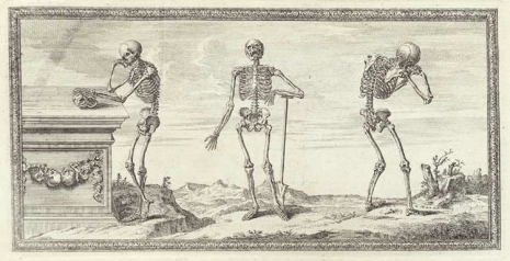
For the Osteographia, Cheselden employed two artists, Gerard Vandergucht and Jacob Schijnvoet, to illustrate the anatomy of bones. To ensure accuracy in the illustrations, Cheselden made use of a camera obscura which transposed the image of each bone onto paper for the artists to copy. However, many of the skeletons were presented in strange so-called realistic positions—for example the skeleton of a cat arching its back at the sight of an approaching dog, or a man kneeling down praying. This was achieved by wiring the skeletons into position, which more often than not detracted from any attempt at factual representation. Thus the book proved to be a failure, though today Cheselden’s Osteographia is considered one of the great historical works of art and science.

Those with an interest can view the whole book here.
 
001bonesskeletons1.jpg
 
002bonesskeletons2.jpg
 
More of dem bones, after the jump…
 

READ ON
Posted by Paul Gallagher
|
06.05.2017
10:43 am
|
Blood and Guts in High School: Beautiful and surreal illustrations for science text books

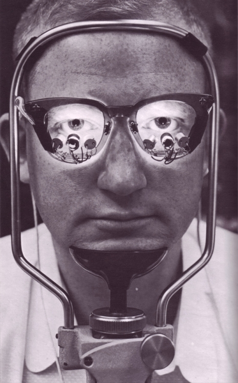
002Etude-des-mouvements-de-loeil.jpg
 
From what I can gather Le Livre de la Sante or The Book of Health or the Encyclopedia of Mind, Body and Health by Joseph Handler was a multi-volume series of text books on science, anatomy, biology, psychology and health intended for use in the classroom. Reading these text books must have been a blast as page after page is filled with the most beautiful day-glo colored illustrations by an incredibly diverse range of artists and graphic designers.

Published in Monte Carlo between 1967 and 1969, Le Livre de la Sante was also made available in an Iranian edition—which kinda shows how hip Iran was back then. Handler’s educational books are still available to buy—and 50 Watts has uploaded a whole library of pages from these books which can be viewed here.
 
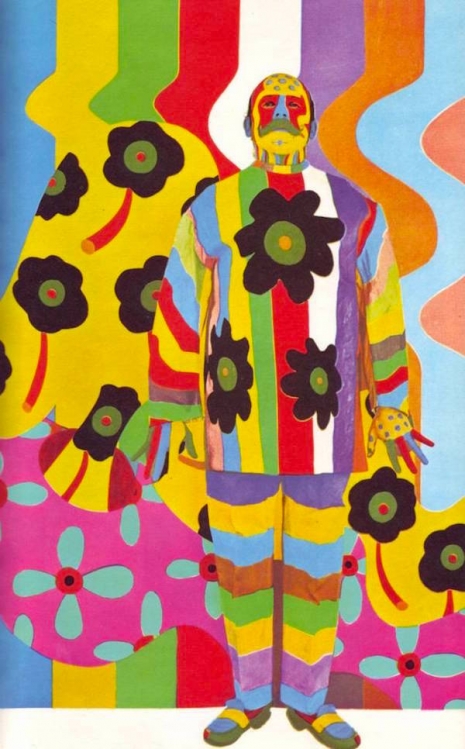
001Lhomme-tableau-tableau-de-Pinoncelli-photo-Ito-Josue.jpg
‘L’homme tableau de Pinoncelli’ by Josue.
 
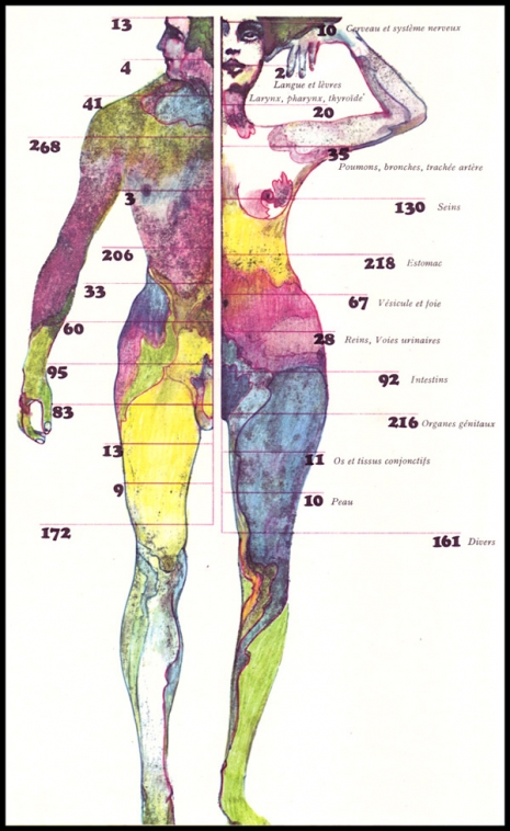
016Le_repartition_des_cancers_Osterwalder.jpg
‘Le repartition des cancers’ (the distribution of cancers) by Osterwalder.
 
More exquisite illustrations from ‘Le Livre de la Sante,’ after the jump….
 

READ ON
Posted by Paul Gallagher
|
11.14.2016
12:03 pm
|
Soft Machine: Body part sculptures made from fabric
08.10.2016
11:31 am
Topics:
Tags:

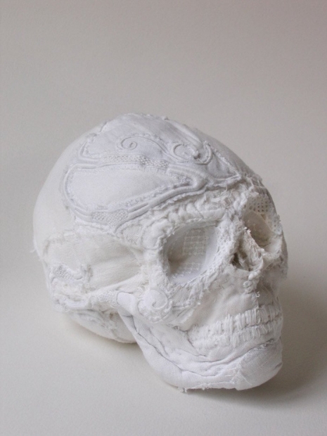
001karinjol001.jpg
 
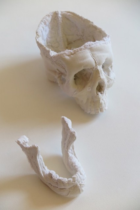
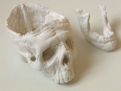
Karine Jollet is an artist whose work explores the enigma of the human body. She creates “soft sculptures” out of recycled fabrics into anatomically correct body shapes.

Jollet sees an analogy between her working materials and “our own biological tissues: bones, fibers, crystals.”

I start with old bed sheets and shirts, embroidered handkerchieves and second-hand fabrics that I cut up, put the fragments together, pad them and then sew them by hand.

The resulting anatomical sculptures are beautiful yet slightly surreal—a lace skull, a pillow brain, squeezable metatarsals. Jollet believes anatomy possesses “a hidden dimension that connects us to an invisible, ideal order of things and also to a secret, dreamlike reality.”

These soft sculptures are produced in a dazzling white as “white evokes a world beyond the visible, beyond the living space of unity and purity.”

More of her work can be viewed here.
 
002Akarjol002A.jpg
 
002Bkarjol2B.jpg
 
More cloth body parts, after the jump…

READ ON
Posted by Paul Gallagher
|
08.10.2016
11:31 am
|
Disturbingly beautiful (almost dirty) images of human anatomy from the 1700s (NSFW)

0_01platesanat.jpg
 
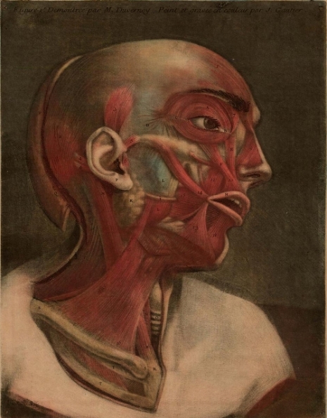
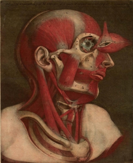
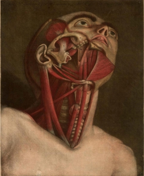
Jacques Fabien Gautier was a printmaker, painter, anatomist and philosopher who is now best remembered for his often lurid anatomical illustrations.

Born in Marseilles in 1716, Gautier began his career as a painter before moving onto printmaking where he developed an interest in the techniques of color printmaking which were then being pioneered by Jacob Christoph Le Blon (1667-1741). Gautier posited the theory colored prints could be created in much the same way as colored patterns were woven into cloth.

In 1736, Gautier moved to Paris—as he believed only great ideas came from great cities. Here he met Louis-Bertrand Castel, a mathematician and scientist who encouraged Gautier to investigate his theories into color printing. However, many of Gautier’s proposals for three and four color printing had been already developed by Le Blon. In 1738, Gautier joined Le Blon’s color-printing workshop but left after only six weeks. He then adopted Le Blon’s ideas and established a printmaking business as a four color printmaker.

Gautier had one good idea—he decided to produce all of the color anatomical illustrations for medical studies. He collaborated with Jacques Francois Duverney, a lecturer in anatomy at the Jardin du Roy. Together they produced l’Essai d’anatomie or Myologie complete en couleur et grandeur naturelle, composée de l’Essai et de la Suite de l’Essai d’anatomie en tableaux imprimés (1746) and Anatomie de la tête, en tableaux imprimés qui représentent au naturel le cerveau sous différentes coupes, la distribution des vaisseaux dans toutes les parties de la tête, les organes des sens et une partie de la névrologie, d’après les pièces disséquées et préparées par M. Duverney, en 8 grandes planches dessinées, peintes, gravées et imprimées en couleur et grandeur naturelle, par le sieur Gautier or Anatomie de la tête (1748).

The collaboration earned Gautier respect. He became known as a philosopher and anatomist became and was made a member of the Dijon Academy of Sciences.  In 1752, he published a baffling critique on Sir Isaac Newton’s theory of color—Chroa-génésie—in which amongst other things he claimed:

...the sun as the universal agent and motive force. According to Gautier’s theory, the force of its rays generates planetary motion, and it is the source of light and fire, substances with broad significance and many uses according to his system. Modified, they create thunder, lightening, and such geologic phenomena as volcanoes and earthquakes…

Gautier’s theories showed his “understanding of geometry is even less exact than his understanding of Newtonian optics.” His writing was described as “convoluted” and “unintelligible.” Surprisingly, this did not stop Gautier from being taken seriously (if only briefly) as a philosopher—enough to have the great writer Goethe suggest his treatise on color deserved an answer. Goethe also described Gautier as “an active, quick, rather impulsive man, certainly gifted but more than befittingly aggressive and sensational.”

Some critics considered Gautier veered more towards the sensationalist than the scientific:

[Gautier’s] anatomical illustrations while they may perhaps be fascinating to the layman…impress the critical observer with their arrogance and charlatanery and do not recommend themselves to the student of anatomy either for their faithfulness or their technique.

His later work in particular—when Gautier was acting as both anatomist and illustrator—has been dismissed as:

“....probably aimed at more prurient-minded lay persons than at anatomists.”

In a pre-Bettie Page world, I suppose that you took what you were offered?

Now largely forgotten as a natural philosopher and anatomist, Gautier (or Gautier d’Agoty as he later called himself) is now best known for his illustrative work for Jacques Francois Duverney’s three volumes on anatomy.

A copy of Essai d’Anatomie can be viewed and downloaded here.
 
From ‘l’Essai d’anatomie’.
 
0_02platesanat.jpg
 
0_03platesanat.jpg
 
0_02platesanat.jpg
 
More after the jump…

READ ON
Posted by Paul Gallagher
|
06.03.2016
10:02 am
|
Cuddly and gross knitted dissection specimens
10.05.2015
10:23 am
Topics:
Tags:


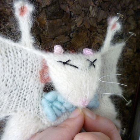
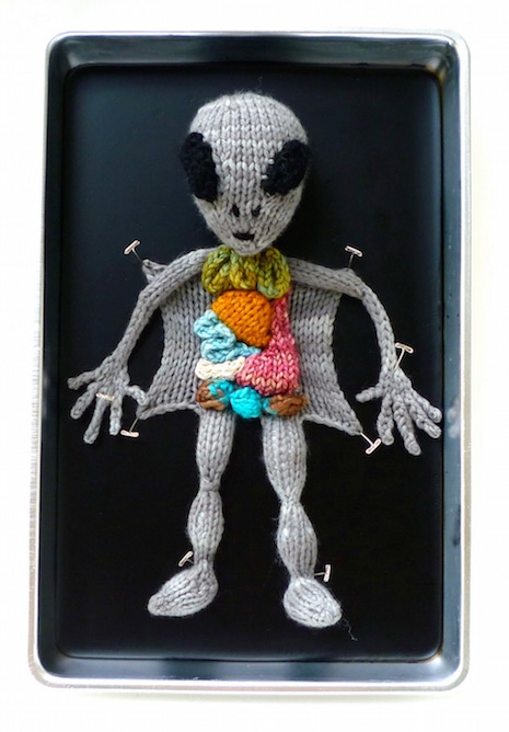
Emily Stoneking says, “If my hands aren’t busy, I’m not happy.” Currently studying German and History at the University of Vermont, Stoneking has an Etsy store featuring crocheted jar cozies and knitted whimsical anatomical studies has allowed her “the freedom to not work for someone else full time, so I can attend school.”

Here we can see Stoneking’s knitted versions of dissected frog, lab rat, earthworm, little alien dude, fetal pig as well as two anatomical studies.


More after the jump…

READ ON
Posted by Martin Schneider
|
10.05.2015
10:23 am
|